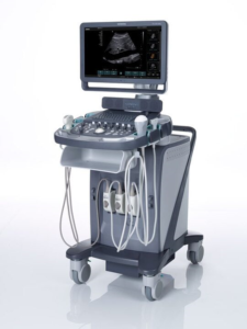
Ultrasound scanners consist of a console containing a computer, a video display screen and a transducer that is used to do the scanning. The transducer is a small hand-held device that resembles a microphone, attached to the scanner by a cord. Some exams may use different transducers (with different capabilities) during a single exam. The transducer sends out high-frequency sound waves (that the human ear cannot hear) into the body and then listens for the returning echoes from the tissues in the body. The principles are similar to sonar used by boats and submarines.
How does the procedure work?
Ultrasound is used to detect changes in appearance, size or contour of organs, tissues, and vessels or to detect abnormal masses, such as tumors.
In an ultrasound examination, a transducer both sends the sound waves into the body and receives the echoing waves. When the transducer is pressed against the skin, it directs small pulses of inaudible, high-frequency sound waves into the body. As the sound waves bounce off internal organs, fluids and tissues, the sensitive receiver in the transducer record tiny changes in the sound’s pitch and direction. These signature waves are instantly measured and displayed by a computer, which in turn creates a real-time picture on the monitor. One or more frames of the moving pictures are typically captured as still images. Short video loops of the images may also be saved.
Doppler ultrasound, a special application of ultrasound, measures the direction and speed of blood cells as they move through vessels. The movement of blood cells causes a change in pitch of the reflected sound waves (called the Doppler effect). A computer collects and processes the sounds and creates graphs or color pictures that represent the flow of blood through the blood vessels.